Unlocking the Secrets of Ancient Egyptian Priests Through Modern Technology!
Radiologists at Keck Medicine of USC have turned the lens of modern medicine on history, using high-resolution CT scanning to examine two ancient Egyptian mummies. The subjects, priests Nes Min (circa 330 BCE) and Nes Hor (circa 190 BCE), had been preserved for over two millennia. Remarkably, each body was scanned while still resting inside the lower half of its heavy sarcophagus, which weighed around 200 pounds.
The CT scanner captured hundreds of thin cross-sectional images of the mummies, which specialists then combined into detailed three-dimensional digital models. These reconstructions revealed astonishingly fine details, from facial bone structure to eyelids and lips, giving researchers a clearer sense of the priests’ individual appearances rather than just their shrouded forms.
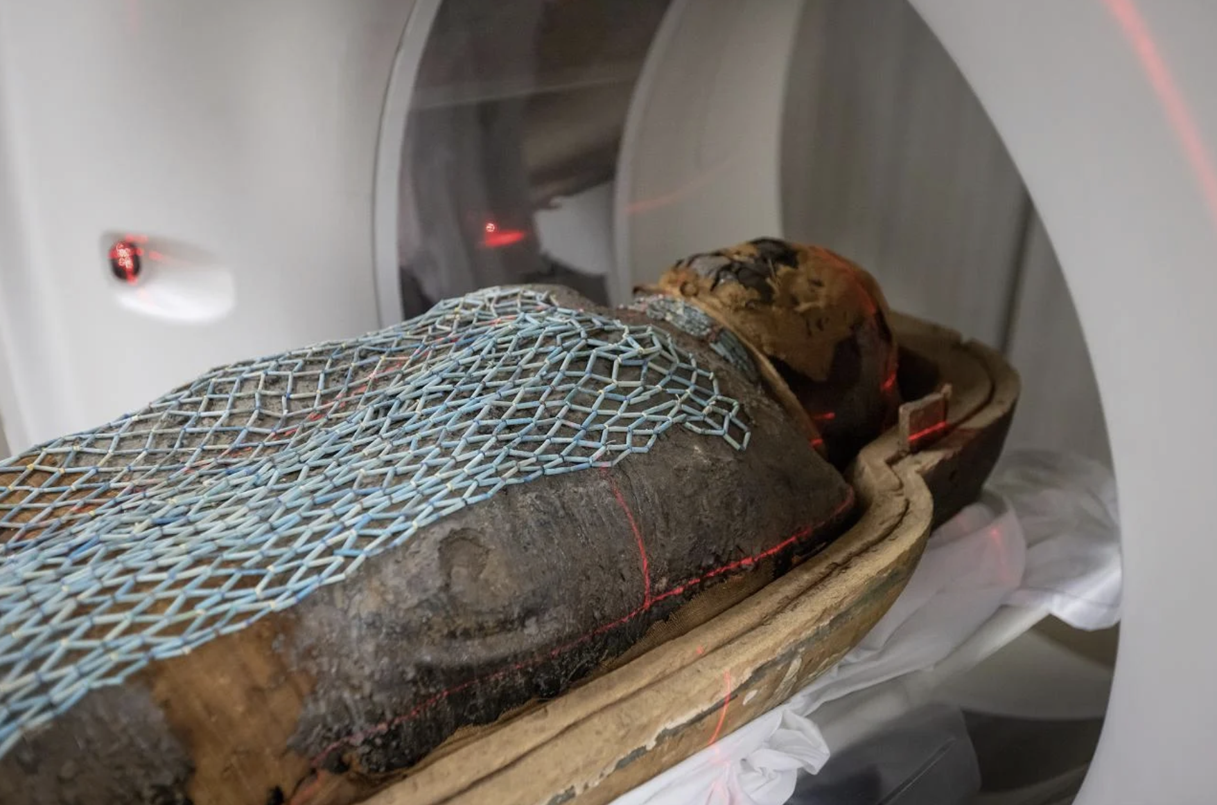
Photo Credit: Ricardo Carrasco III
The scans also offered insights into their health and ageing. Nes Min’s lower spine showed signs of wear and damage, including a collapsed lumbar vertebra, a pattern similar to what modern medicine observes in older adults with chronic back pain. Nes Hor, meanwhile, had severe dental decay and advanced deterioration in a hip joint, suggesting he lived to an older age than Nes Min and may have experienced limited mobility in later life.
Burial items, long hidden within the wrappings, were visible in the CT images. Nes Min, for example, rested alongside small objects shaped like scarab beetles and a fish. The scans allowed researchers to study and measure these items without disturbing the delicate remains, preserving them for future generations.
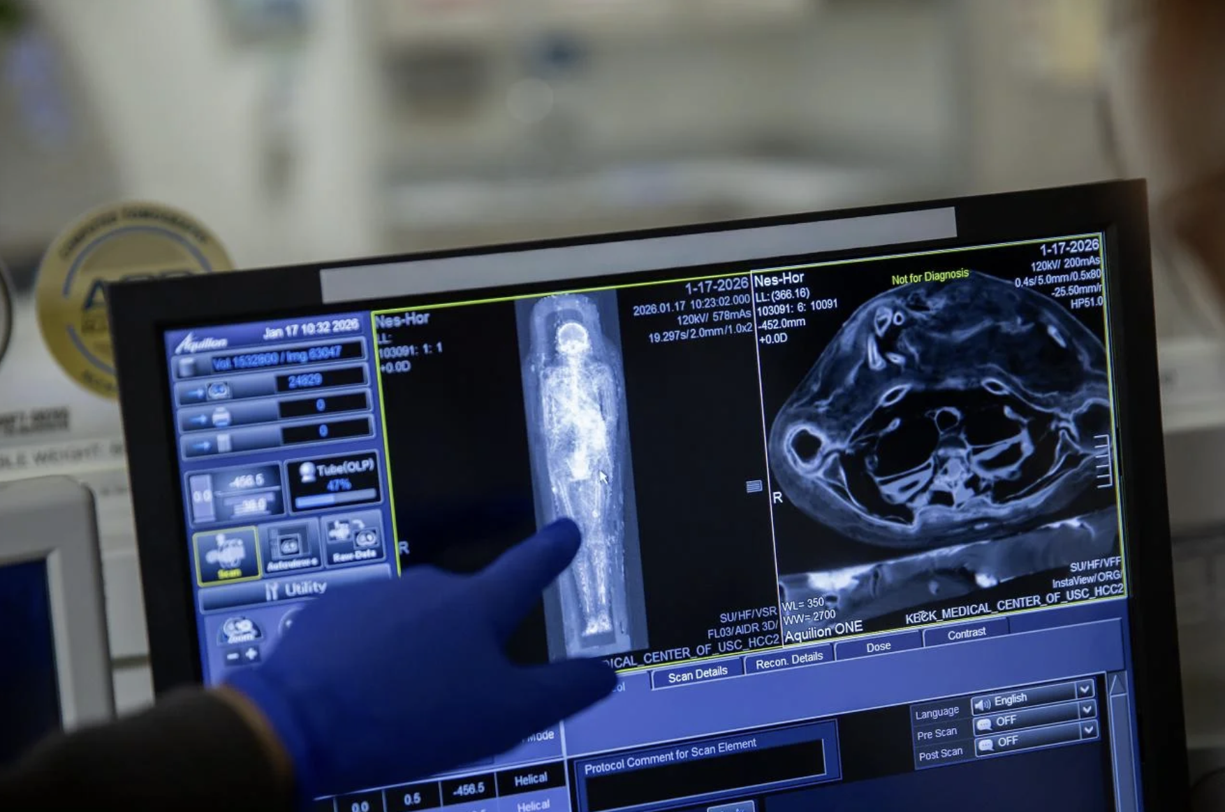
Photo Credit: Ricardo Carrasco III
Following the scans, the team created digital reconstructions of the skeletons and selected burial objects, then produced life-size prints using medical-grade 3D printers. Visitors to the California Science Centre exhibition Mummies of the World, opening on 7 February, will be able to see these replicas alongside the original mummies and digital displays, providing an immersive look at ancient Egyptian life.
This workflow mirrors practices in contemporary medicine, where CT and MRI scans are used to generate 3D models of organs such as the heart, liver, or pelvis. Surgeons use these models to plan complex procedures, select implant sizes, and rehearse intricate steps. Physical prints also help patients understand their own anatomy, allowing for clearer discussions around planned treatments.
By applying these techniques to ancient remains, the mummy project demonstrates how medical imaging not only aids healthcare today but also preserves history. Detailed scans allow researchers to study fragile specimens without damage, offering insights into health, injury, and daily life in ancient Egypt, thousands of years after Nes Min and Nes Hor first lived.